

Private cardiology clinics in Ashford, Margate, Maidstone and South London
Accessible care from expert cardiologists
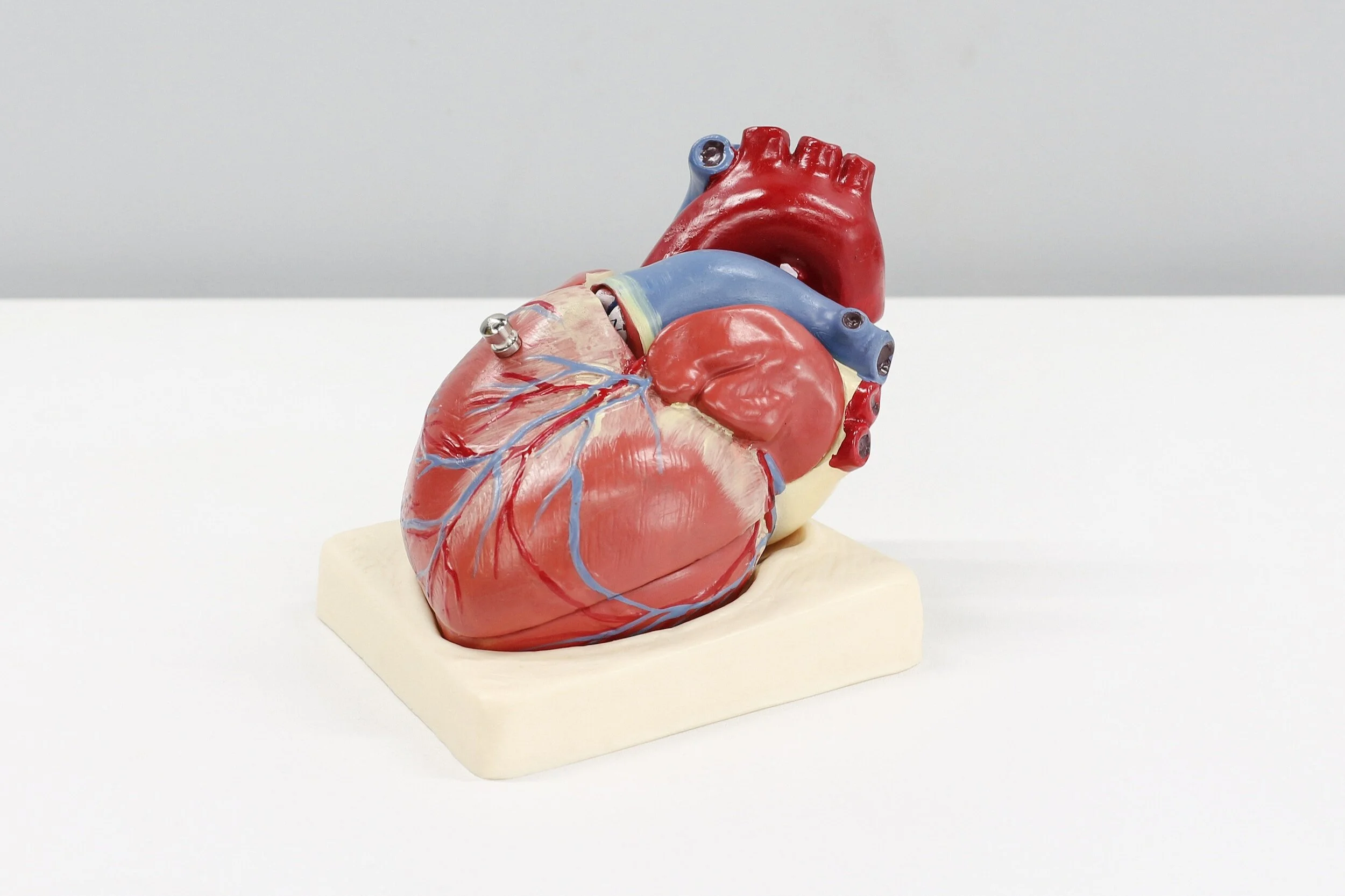
We are a multi-consultant private cardiology practice covering the full range of heart disease. We have particular expertise in chest pain, arrhythmia, heart failure, syncope, breathlessness, autonomic dysfunction and PoTS. With highly qualified consultants in clinics across Kent and London there is no need to compromise on quality or on convenience.
-

Private Appointments
We see insured and self-pay patients with all types of heart disease. We do not require a GP referral. First appointments last 40 minutes to allow your heart specialist to reach an in-depth understanding of your cardiac issues.
-

Diagnostic Tests
With our local knowledge we use only the best private hospitals and most qualified staff for cardiac tests such as echocardiography, ECG, Holter, ambulatory BP, cardiac MRI, angiography and tilt table testing. All investigations are available promptly at private hospitals close to you.
-

Cardiac Procedures
We will support you through your whole journey with a new heart problem including diagnostic procedures such as coronary angiography and electrophysiological (EP) studies, as well as ablations, pacemaker and defibrillator implants.
Request a call-back to discuss a consultation

Our London heart clinics
Our regular cardiology clinics in London are held at:
Guthrie Clinic at Kings College Hospital
The Syon Clinic in Brentford

Our consultant cardiologists

Dr Idris Harding


“I am passionate about cardiology and heart rhythm care. All my patients should feel they’ve had the best possible treatment.”
Dr Idris Harding
Consultant Cardiologist and Cardiac Electrophysiologist

Dr Suzan Hatipoglu
“As a specialist in cardio-oncology my goal is preventing, diagnosing, and treating heart complications that can arise from cancer treatments.”
Dr Suzan Hatipoglu
Consultant Cardiologist and Cardiac Imaging Specialist
Our clinical nurse specialist

Mrs Sjouke Ashton-Clarkson
“Heart failure can be a lonely and scary experience but modern treatments can be transformative.”
Sjouke Ashton-Clarkson
Senior Cardiac Nurse Specialist and Nurse Prescriber
Praise from our patients
“Dr Harding is a compassionate, friendly
and informative consultant.”
Arrhythmia patient, Jan 2024
“Dr Hatipoglu is very authoritative and clear. I have confidence in her expertise.”
Heart failure patient, Jan 2025
“I finally feel that I have been heard. Dr Harding is professional, empathetic and transparent.”
PoTS patient, Dec 2023


Learn about cardiac symptoms
-
There are many causes of pains in the chest but the most concerning we see in our heart clinic is coronary heart disease, which can cause angina and heart attack.
The pain of heart disease is usually described as tight or heavy in nature and may spread to the arms and/or jaw. It may be accompanied by other features such as shortness of breath, dizziness, or profuse sweating. However the pain of heart disease varies considerably between individuals and just because pain doesn’t fit the pattern described this does not rule out heart disease as the cause.
NHS advice is to treat any pain in the chest lasting for more than 15-20 minutes as a potential heart attack and to call 999.
Aside from coronary heart disease, other cardiac causes of chest pains include aortic dissection (a serious problem with the main blood vessel leading out of the heart) and pericarditis (an inflammatory condition affecting the lining of the heart).
There is also a long list of non-cardiac conditions that can cause chest pain.
-
Shortness of breath or “dyspnoea” is most often caused by diseases of the heart or lungs but other conditions (for example anaemia and thyroid disorders) may also contribute.
For this reason, assessment of breathlessness usually starts with a full examination and some blood tests to rule certain causes in or out.
Heart diseases that cause breathlessness include coronary heart disease (angina), heart failure and some heart rhythm abnormalities (arrhythmias).
Angina classically causes pain,. but some people (especially older patients, and patients with diabetes) perceive the pain as a sensation of breathlessness.
Some patterns of breathlessness strongly suggest a particular cause, for example breathlessness that comes on at a predictable level of exertion - at the same point walking up a particular hill, say - and goes off rapidly with rest, may be suggestive of angina caused by narrowing of the heart arteries.
Tests or investigations that may be used to uncover the cause of breathlessness include lung function tests and chest X-ray to look at the structure and function of the lungs, as well as an echocardiogram and ECG to examine the structure and function of the heart. In some cases, extended ECG monitoring, either using ambulatory ECG monitors (sometimes called “Holter monitors”) or exercise ECG may be used to look for heart rhythm problems that may be intermittent.
As well as exercise ECG, various other cardiac stress tests can be used to diagnose whether breathlessness is due to angina. These tests include stress echo, stress nuclear scans and stress MRI of the heart. Each cardiac stress test has its particular advantages and drawbacks and choosing the right test for the right person is part of the skill of cardiology.
If a stress test does suggest that the cause of breathlessness is ischaemic heart disease (i.e. a blood supply problem within the heart) then the next step is generally an assessment of the coronary arteries with a CT coronary angiogram scan or invasive coronary angiogram to look for narrowings in the large arteries on the surface of the heart.
-
The symptom of “palpitations” means an abnormal awareness of the heart beat. However this is a difficult thing to describe and in the heart clinic, people may use many different terms to describe the feelings associated with abnormal or racing heart beats. One person’s “flutter” is another’s “skipped beat”, “sinking feeling”. or “heart stop”. The terminology used really is endless and while some of it is very descriptive, only occasionally does the description of the palpitations themselves lead to a diagnosis without the need for further tests.
There are many different heart rhythm abnormalities ranging from one-off extra beats (“ectopics”) through to persistent and chaotic rhythms such as atrial fibrillation. While some arrhythmias carry an excellent prognosis and therefore have very little significance, others do have implications for the sufferer’s health. For example, ectopic beats, if they occur repetitively and frequently, can impair the heart’s pump function over time, and in some rare cases can lead to heart failure. This is not common, but to rule out the possibility, further tests such as ambulatory ECG for 24, 48 or 72hrs (sometimes called “Holter monitors”) plus an echocardiogram or cardiac MRI, are often needed.
The gold standard for diagnosing the cause of palpitations is to make a recording of the heart’s electrical activity while the palpitations are happening. Until recently the only way of doing this was with increasing lengths of ambulatory monitoring, but devices such as Apple Watch, FitBit, Kardia Alivecor, Withings Move ECG and others have revolutionised this area. Most modern electrophysiologists (heart rhythm specialists) have integrated all these new technologies into their clinics.
Atrial fibrillation is the most common cause of palpitations and increases with age. This disease can be associated with a risk of strokes in some people, so making a diagnosis of this condition can be quite important, as treatments are available to reduce the risk of stroke in some people.
-
It is important to distinguish between dizziness or lightheadedness, versus vertigo, which is a sensation of spinning. Vertigo is typically caused by brain or ear problems, whereas dizziness and lightheadedness have a much wider range of potential causes, including disorders of the heart and circulation.
Anything that causes the blood pressure to drop can cause lightheadedness, and in extreme cases, loss of consciousness. Low blood pressure can result from dehydration, position changes, changes in blood vessel responsiveness, or changes in the heart’s ability to pump blood through the circulation.
There is a long list of diseases that can cause low blood pressure, and most cardiologists will approach the issue first with a thorough history and clinical examination to work out if there is a pattern to when the blood pressure drops and whether there is any obvious and severe cardiac cause, for example valve disease such as aortic stenosis.
Further investigation is guided by the initial assessment and may include provocation tests such as tilt table and active stand tests, which can trigger blood pressure drops in patients with orthostatic hypotension, reflex syncope or PoTS.
Sporadic and unpredictable blood pressure drops may result from changes in heart rate or rhythm. These may be diagnosed using prolonged heart rhythm monitoring via devices such as Holter monitors and implantable loop recorders. If the symptoms are not severe and disabling then heart rhythm monitoring via a consumer wearable device such as an Apple Watch, Kardia AliveCor, FitBit, Withings Move ECG or similar device may be possible.
-
Fluid retention leading to swollen legs and abdomen is a common symptom of many medical conditions. Many people also experience mild swelling of the lower limbs after prolonged standing, or during hot weather. A key difference is that “normal” lower limb swelling tends to go down overnight and is not “progressive”; i.e. it does not get worse day after day.
In a heart clinic, progressive fluid retention is often a sign of heart failure, a condition where the heart does not pump enough blood to meet the demands of the body. In this condition, weakness of the heart causes changes in the kidneys leading to fluid retention and ultimately to swollen limbs and weight gain as water is retained in the body instead of being passed as urine.
Other conditions can mimic the fluid retention of heart failure. A typical workup for oedema (the medical word for fluid retention) will include blood tests to make sure that the fluid retention is not due to an underlying kidney or liver problem. In general practice, a blood test called NT-proBNP is used to give a rough idea of whether the heart is pumping effectively or not.
Once kidney and liver disease is ruled out, or if the NT-proBNP level is high, then an echocardiogram or cardiac MRI scan may be used to find out in what way the heart has become weak. Additional cardiac tests such as ECG and ambulatory ECG may also be done to find out whether the heart rhythm is contributing to the weakness of the heart.
Most new diagnoses of heart failure are due to previous heart attacks or angina weakening the heart muscle over time, but other causes include acquired or inherited weakness of the heart muscle - a condition called cardiomyopathy.
Treatments for heart failure differ depending on what the underlying cause is. Cases of heart failure due to an abnormal rhythm may require implantation of a pacemaker, or ablation procedures to return the heart rhythm to normal.
If treatment with medication fails to improve cardiac function and symptoms then selected patients may benefit from implantation of a cardiac resynchronisation device or defibrillator.
-
PoTS is a term that describes persistent heart racing on standing up, sometimes accompanied by symptoms of low blood pressure (dizziness or lightheadedness) and fatigue.
PoTS has a range of causes and can be diagnosed in association with a number of different underlying illnesses. PoTS-like symptoms have also been described as part of Long COVID.
Wearable technology
We take full advantage of the tech you are already carrying on your wrist or in your pocket to diagnose your heart problems.
Even if you don’t have an ECG-capable device it may still be possible to install an app on your smartphone to diagnose atrial fibrillation using your device camera, for example.

Our Secure Patient Portal

Access all your clinic correspondence and investigations via our secure online portal.
Download clinic letters and review test results at your convenience.