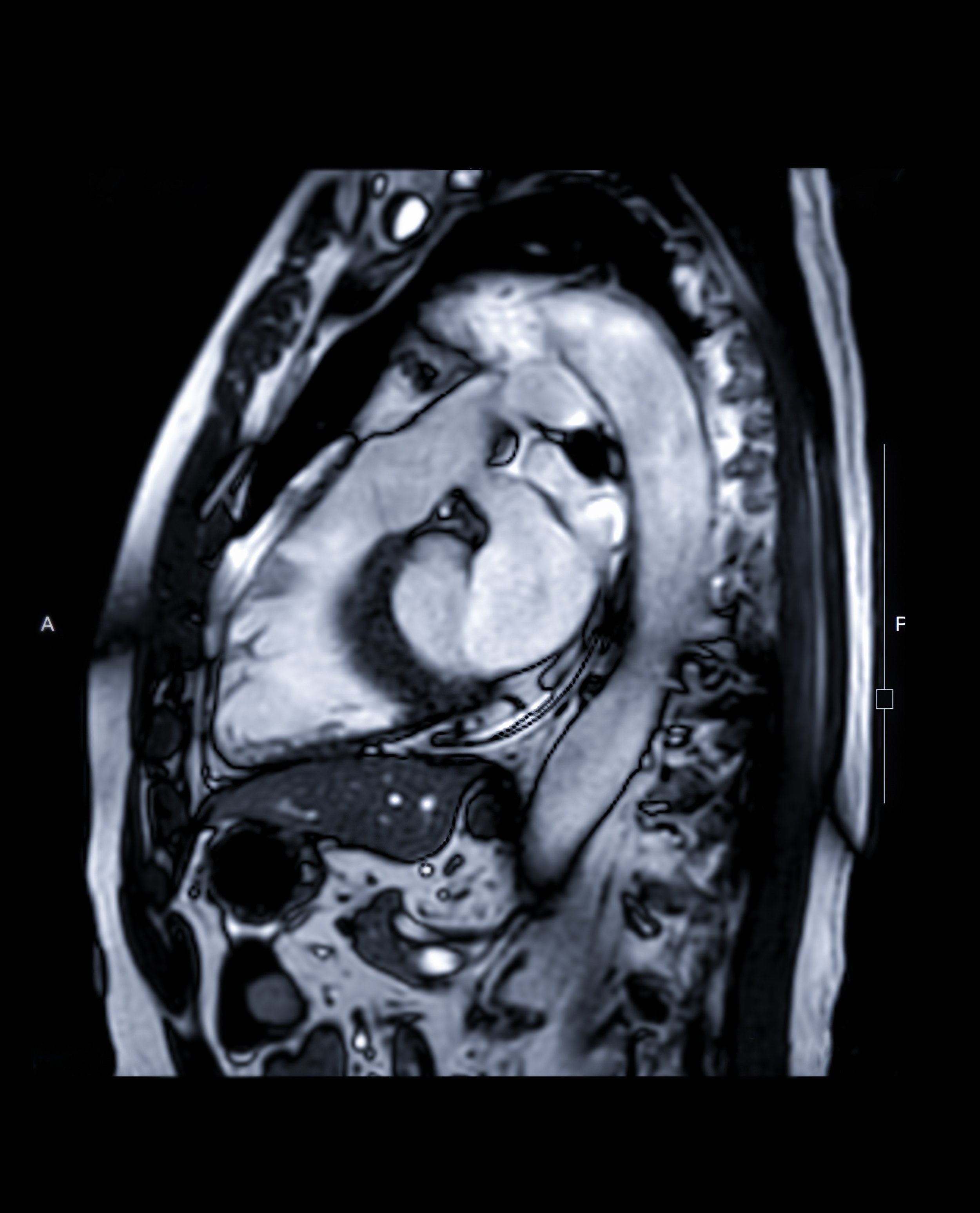
MRI scans for heart disease
MRI uses electromagnetic imaging to give pictures and videos of the beating heart that are used to diagnose many cardiac conditions.
Cardiac MRI
A basic MRI scan of the hear takes half an hour to 40 minutes during which the patient should lie quite still and follow breathing instructions. The scanners are often confined spaces and fairly noisy due to the high speed that magentic coils must move at the obtain images of the heart.
MRI scans are very safe. The only real exception to this is for patients with metalwork in or around the body: an MRI scanner causes any metal in the surrounding area to move violently. Patients with ferrous metal implants, including some older joint replacements, etc, cannot have MRI scans for this reason. Likewise patients who have been injured with metal fragments where there is a chance that this metal could still be in the body should not have MRI.
A basic MRI scan will provide information about the size and motion of cardiac structures including the heart chambers, their walls, and heart valves. This information can be used to diagnose heart failure, heart valve disease and abnormal cardiac anatomy such as septal defects (holes in the heart walls).
MRI with contrast
As with other scans (CT and echo, for example) injections can be given during an MRI scan to hilight certain structures. Commonly, Gadolidium contrast is used during MRI scans. This dense substance highlights the presence of scar and abnormal tissue within the heart muscle. This is information that is not readily available by any other technique, and may be used to inform decisions about stenting procedures, or about whether a patient should have a defibrillator.
Stress MRI scans
Just like stress echo, and exercise ECG, MRI imaging can also be used to image the heart during high workload. This can give information about the blood supply to the heart and how well heart muscle and heart valves work under conditions of stress.
This information can be used to assess whether stenting procedures, or other surgical interventions such as valve operations, are needed.
Who can’t have an MRI?
MRI scanners are generally quite enclosed and noisy places. Headphones and music are routinely used as distractions during a scan. Most MRI departments are highly skilled at helping patients relax and get through a scan, but patients with severe claustrophobia or anxiety may struggle to complete a scan.
Patients with ferrous (iron-containing) metal inside the body cannot have an MRI scan due to the risk of the metalwork moving and causing injury. For this reason, most medical and surgical implants are now manufactured with non-ferrous metals.
Pacemakers and MRI
For many years, due concerns about heating the metalwork and causing the devices to malfunction, doctors avoided putting patients with cardiac defibrillators and pacemakers into MRI scanners. The safety rules on this are changing, however, as it is increasingly recognised that - with appropriate safeguards - most pacemakers and defibrillators can be safely scanned.
This is not universal, however, and patients with cardiac devices, or any other metalwork or metal debris in the body must ALWAYS tell MRI staff about this, BEFORE getting anywhere near the MRI scanner. Serious injury or death could result from failure to disclose this vital information.

